Inside WashU Medicine highlights the people, programs, departments and images that define WashU Medicine. This image gallery features a selection of visuals from WashU Medicine investigators. Spanning art and science, these images represent research in eye development, fertility, lung disease, neuroscience and bone health.
To submit scientific images, photography, events or story ideas for consideration for future issues of Inside WashU Medicine, visit our submissions page.
Developing eye
Winning image of the 2026 Science in Focus: Art Beyond the Microscope competition in the John F. Hardesty, MD Department of Ophthalmology & Visual Sciences
At embryonic day 14.5, the developing mouse eye reveals the emerging organization of the lens, retina and vitreous humor, offering a glimpse into eye development.
Image credit: Pooja Rathaur, PhD, from the lab of Steven Bassnett, PhD, in ophthalmology & visual sciences
Transparent ovary
A method called “tissue clearing” makes mouse embryonic ovaries transparent. Combined with fluorescent cellular staining and high-resolution confocal imaging, this method reveals the number of eggs (oocytes) in the whole ovary, along with their shapes and distribution patterns.
The lab of Helen McNeill, PhD, in the Edward Mallinckrodt Department of Developmental Biology is using these techniques to answer questions about fertility.
Movie credit: Ling Zhang, PhD
Bone cell communication
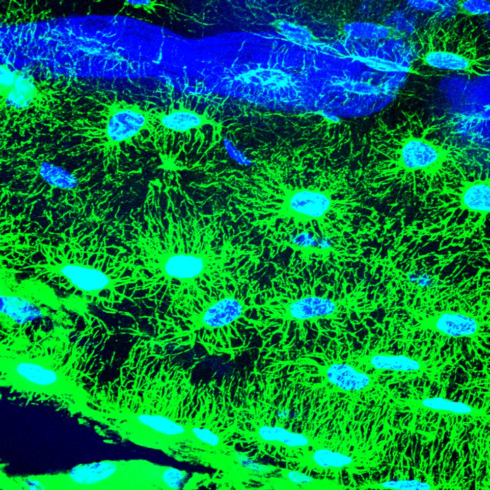
Osteocytes are cells that maintain mature bone tissue. Green staining shows the dendrites that osteocytes produce to communicate with one another and other cell types.
Researchers in the lab of David Ornitz, MD, PhD, in developmental biology have identified key signals important for the formation and maintenance of healthy osteocytes, which advances efforts to understand and prevent bone loss and osteoporosis as people age.
Image credit: Debabrata Patra, PhD
Newt lens
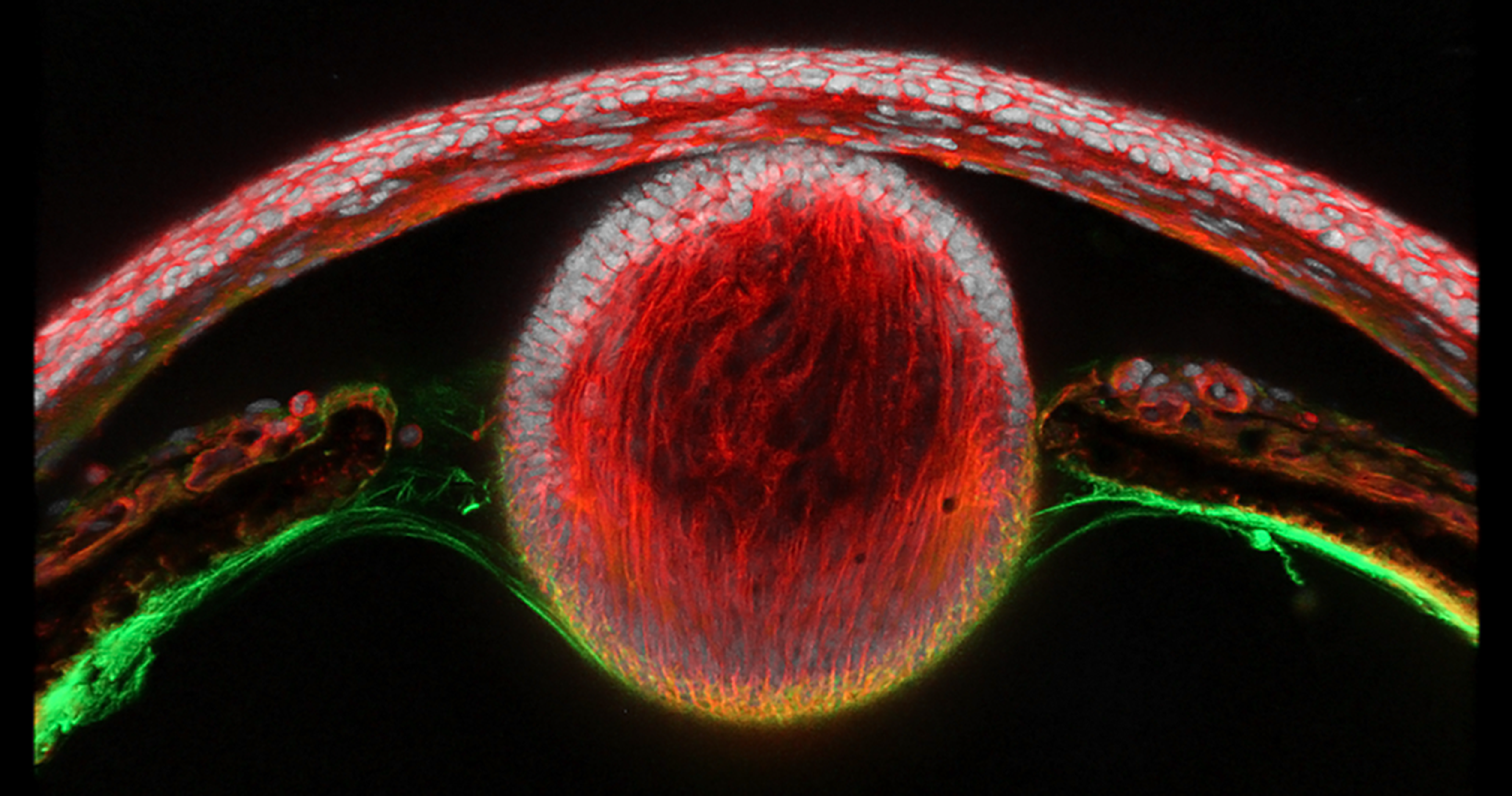
Finalist in the 2026 Science in Focus: Art Beyond the Microscope competition
Thirty days after removal of the newt lens, a new lens (the central red sphere) has regenerated from the inner margin of the iris. Transdifferentiation — the transformation of one differentiated tissue type into another — is a property of certain amphibians and was first documented in the eye by Charles Bonnet in 1781.
Image credit: Pooja Rathaur, PhD, and Konstantinos Sousounis, PhD, from the lab of Steven Bassnett, PhD, in ophthalmology & visual sciences
Reprogrammed neurons
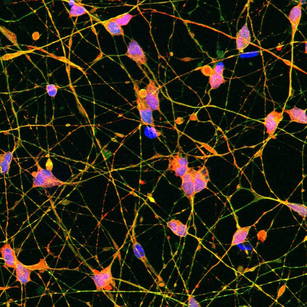
In the lab of Andrew Yoo, PhD, in developmental biology, researchers have developed a way to reprogram human skin cells into different types of neurons. The technique allows them to study the effects of age on neurons — which retain the age of the person who donated the skin sample — and how that affects the development of neurodegenerative diseases, such as Huntington’s and Alzheimer’s.
In a recent study, Yoo’s team used reprogrammed human neurons (pictured) to investigate the effects of aging on mitochondria, the energy factories of cells.
Image credit: Eva Klinman, MD, PhD, a WashU Medicine neurologist who was a 2025 STAT Wunderkind
Eye inflammation
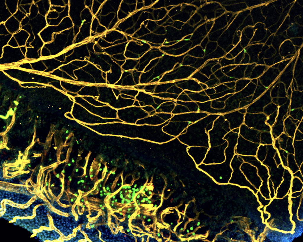
Finalist in the 2026 Science in Focus: Art Beyond the Microscope competition
Immune cells (green) from patients with eye inflammation, called uveitis, infiltrate the retina. This is a hallmark of intermediate uveitis (which occurs behind the lens of the eye), posterior uveitis (in the back of the eye) and panuveitis (throughout the eye).
Image credit: Codie Cook, technician, from the lab of James Walsh, MD, PhD, in ophthalmology & visual sciences
Infected lung
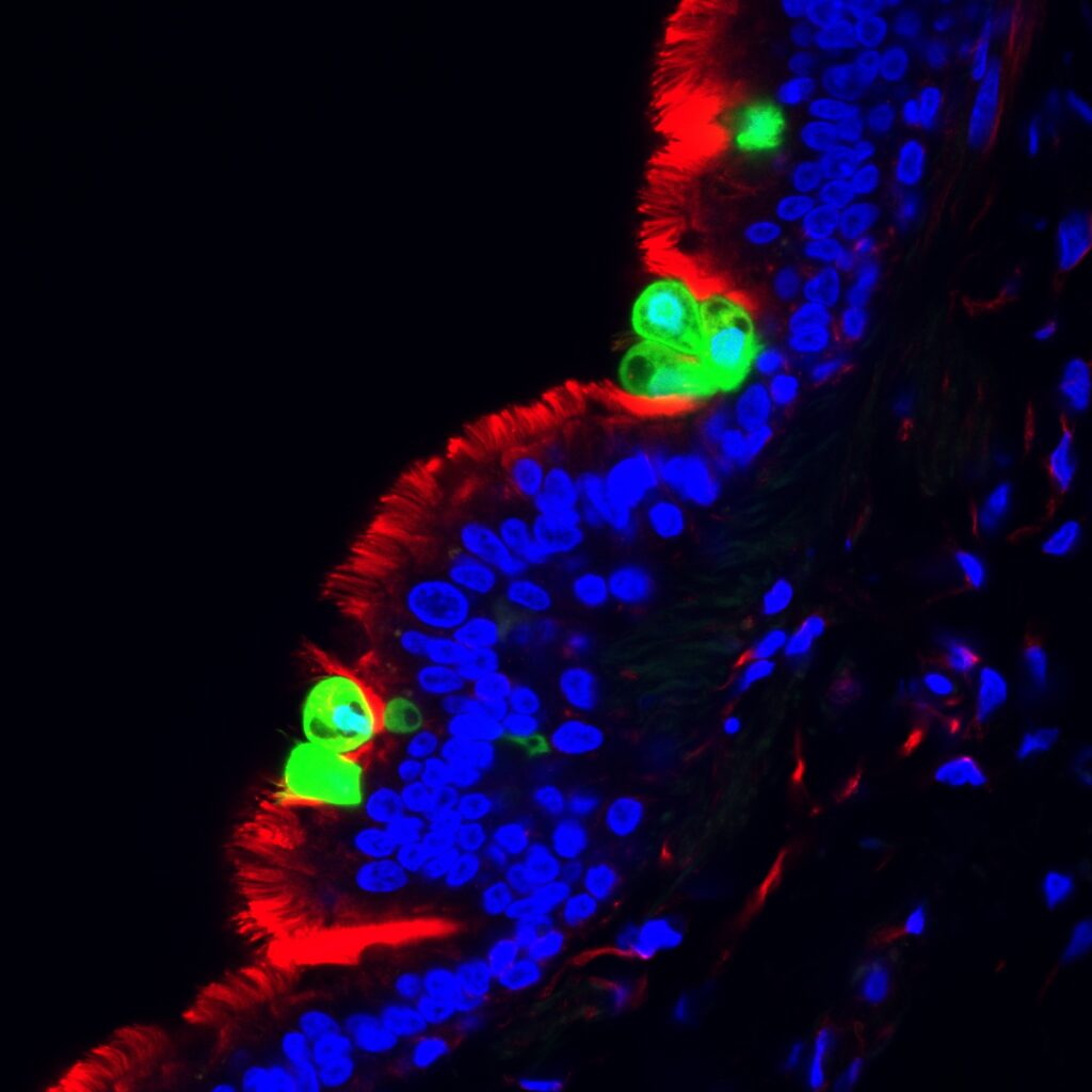
Researchers in the lab of Xingbin Ai, PhD, in the John T. Milliken Department of Medicine’s Division of Pulmonary & Critical Care Medicine, study the progression of lung diseases, including viral infections such as respiratory syncytial virus (RSV).
Shown is a slice of a human infant lung infected with RSV (green). Cilia (red) are tiny hair-like structures on the surface of cells that help form a protective barrier and keep the lungs clear. In infants, RSV is much more damaging to lung cilia than it is in adults, and the Ai Lab is investigating why.
Image credit: Caiqi Zhao, PhD
Uterine lining
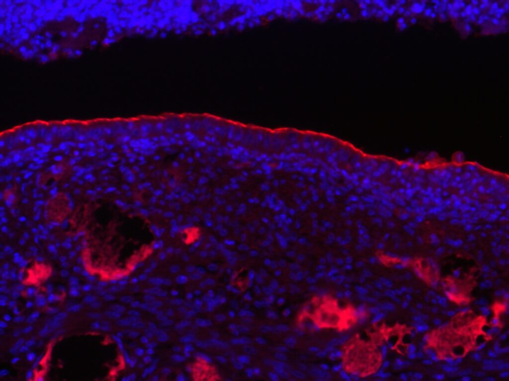
Liang Ma, PhD, and his colleagues in the Division of Dermatology are studying the uterine lining with a focus on infertility.
Shown is the uterine lining of a mouse that lacks key retinoic acid signals needed for the uterus to become ready for an embryo to attach. The team found that without these signals, a protein barrier on the uterine surface remains in place (red line), which may help explain some forms of infertility.
Image credit: Yan Yin, PhD